Saturday, April 19, 2008

Unit IV Compendium

Compendium Unit IV
Chapters 16-17
16.1 Human Life Cycle

Human reproduction is possible when a person completes puberty. In girls, that is reached at age 11-13 and boys at 14-16 years of age. To follow are the different functions of organs for each sex:
Male: Produce sperm Female: Produce eggs
Nurture sperm Transport egg to uterus
Penis delivers sperm into vagina Vagina receives sperm and provides conduit to egg
Testes produce sex hormones Uterus provides locale for fetal development
Vagina is pathway for delivery
Ovaries produce sex hormones
There are two types of cell division: mitosis and meiosis. Mitosis is duplication from one cell dividing producing two cells each containing all 46 chromosomes. Meiosis occurs only in the testes and ovaries of the reproductive organs. Each sperm and egg cell produces contain only 23 chromosomes so that when united, the zygote will have all 46 chromosomes.

16.2 Male Reproductive System

The testes (gonads) are the primary sex organs of the male reproductive system. The testes produce sperm and sex hormones. The scrotum will regulate the temperature of the testes by raising or lowering the testes from the body. Too warm and the sperm will not be viable. The sperm than mature and are stored in the epididymides. The sperm then leave the epididymides and can be stored in the vas deferens or transported onward. After entering the ejaculatoroy duct, nutrients and semen are contributed by the seminal vesicles, prostate gland, and bulbourethral glands.
Male reproductive anatomy:


I will not be commenting on the act of intercourse, since this is a public internet site. There may be greater than 400 million sperm in 3.5 mL of semen.
The Seminiferous Tubules are the locations of sperm production. It takes 74 days for sperm to develop. Mature sperm have 3 parts, head, middle piece, and tail. The head is covered by acrosome, which contains an enzyme to penetrate the egg. The middle piece has mitochondria which provides energy for movement. The tail flagellates to propel the sperm. The interstitial cells in the testes produce the sex hormones. The hypothalamus in the brain produces gonadotropin-releasing hormone (GnRH) that stimulates the anterior pituitary to release gonadotropic hormones, both follicle stimulating (stimulates sperm production) and luteinizing hormones (stimulates testosterone production). Testosterone maintains normal development and functioning of the sexual organs. It also gives males some of their male traits, such as greater muscle mass, lower voices, adam’s apple, broader shoulders and greater height.

16.3 Female Reproductive System

Ovaries are the female gonads. The produce eggs, estrogen and progesterone. The ovaries are located at one end of the fallopian tubes. They are not connected. The fallopian tubes have fimbriae (projections that sweep over the ovaries) and cilia which “brush” the eggs after they emerge from the ovaries in the direction of the uterus.


An egg is only viable for 6-24 hours until fertilization occurs. Fertilization usually occurs in the fallopian tube. The developing fetus (zygote, at this stage) arrives and implantation occurs in the thickened uterine lining. The uterus prior to emplantation is approx. 5 cm wide and can stretch to over 30cm wide when pregnant. The lining of the uterus is called the endometrium comprised of two layers, the basal laer and the functional layer. The cervix is at the base of the uterus and leads to the vaginal canal.


The external genitalia is collectively called vulva. The outermost folds are called labia majora. Moving medially, the next folds are called labia minora. Ventrally, they join to form a foreskin for the clitoris, this is the organ of arousal in a woman. Just dorsal to that is the urethra and then the vagina.

16.4 Female Hormone Levels

Hormones cycle an average of every 28 days. This is ovary-driven. An ovary initially contains as many as 2 million follicles and only about 400 will ever mature to produce an egg. Generally speaking, only one egg is produced every month.
Genetic spicing gone wrong. The chicken must have died,
there's not a full dozen.


Back to the basics . . .
The human cycle is depicted in the picture below:
http://www.anselm.edu/homepage/jpitocch/genbio/repronot.html

After the oocyte is released, the follicle develops into a corpus luteum. This produces progesterone and a little estrogen. As in men, the hypothalamus controls sexual function by releasing gonadotropin-releasing hormone. This stimulate the pituitary to produce FSHand LH, which control the ovarian cycle. During the first half of the cycle, the FSH stimulates the follicle in the ovary to produce estrogen which cignals the hypothalamus to secrete GnRH which in turn leads to a surge of LH production by the anterior pituitary and ovulation. The LH matures the corpus luteum and progesterone is secreted. Estrogen is responsible for our feminine traits. In encourages fatty accumulation under the skin, lending a softer appearance. The ovarian cycle will cease at around 50 years of age. The ovaries fail to respond to GhRH and no longer secrete estrogen and progesterone.
The uterine cycle is the response to the fluctuating hormones. The lining of the uterus will thicken to accommodate a possible developing embryo and this will slough off if an egg is not fertilized that month resulting in a menstrual period.
Pregnancy: Once a sperm has fertilized an egg, it becomes a zygote. This implants in the lining of the uterus. At the place where the molecules of fetal blood and maternal blood are exchanged, a placenta will develop. Progesterone production increases and will stop follicles from producing more eggs during the pregnancy. The placenta will begin to produce progesterone and estrogen and the corpus luteum will regress.

16.5 Control of Reproduction

There are multiple methods available to prevent pregnancy from occurring. They are listed below and these rates are applicable only when the methods are used correctly:


Abstinence is the only guaranteed method of birth control and it also has the advantage of no STD transmission. Morning after pills are administered 1-7 days after intercourse has occurred. It upsets the progesterone cycle preventing the implantation of the embryo. RU-486 is a different type of morning-after pill that causes the endometrium to slough, of course the embryo goes with it.
Infertility is deemed such after one year of regularly attempted impregnation. Low sperm count is one cause of infertility. Too much sitting, smoking or drinking alcohol can cause this. Overweight women may fail to ovulate. Blocked oviducts from pelvic inflammatory disease and endometriosis can prevent the journey of the egg to the uterus also causing infertility.
There are technologies available to help infertile couples have children. Artificial insemination is when the sperm is placed in the vagina by a physician. This is helpful when the male has a low sperm count. Sometimes fertility drugs are given to the woman to produce one or more eggs and the sperm is placed directly into the uterus. In Vitro Fertilization, conception is attained in the lab. Then the embryos are transplanted in the uterus of the woman. Sometimes, other women are contracted to give birth. The egg and sperm can be from the desiring parents. Scientists can also inject a single sperm into and egg and then transplant it into the mom’s uterus.

16.6 Sexually Transmitted Diseases

Viral Sexually Transmitted Diseases
HIV – precursor to AIDS. First stage of infection can be asymptomatic, but highly contagious. This may develop into AIDS when the helper T cell count falls dangerously low and the host cannot fight infections. AIDS patients usually die from opportunistic infections that would normally be stopped by a healthy immune system.
Genital Warts – caused by the human papillomaviruses. The carriers may not have warts and may have less noticeable flat lesions. A baby can pick up this virus as it passes through the birth canal. Vaccinations are available for some the common strains of this virus. 90% of all cancers of the cervix are thought to be caused by this virus.
Genital Herpes – Herpes simplex type 2. This is presented with blisters, tingling or itching sensations. Some events may be accompanied with fever, pain with urination, or swollen lymph nodes and the person is very contagious at this time. Newborns may pick this up through the birth canal.
Several types of hepatitis are acquired through sexual transmission. This generally causes liver damage.

Bacterial Sexually Transmitted Diseases

These are curable with antibiotics. Some resistant strains may require taking stronger drugs for a longer amount of time.
Chlamydia – mild or asymptomatic in women. If undiagnosed, can spread from the cervix to the fallopian tubes causing pelvic inflammatory disease, a very painful condition which may result in infertility.
Gonorrhea – can lead to pelvic inflammatory disease in both males and females. Gonorrhea will infect whatever part was exposed to the virus and can be spread to internal parts, causing heart damage or arthritis. Because of increased resistance to antibiotic therapy, 40% of all strains don’t respond to therapy.
Syphilis – also potentially deadly, begins as a small ulcer (chancre) that resolves on its own. Then the infected person will develop a rash that does no itch and is seen on palms and soles of feet. This, too, will resolve. The last stage may affect the cardiovascular system by causing aneurysms and ulcers on the skin or internal organs. Early diagnosis and treatment is vital for successful treatment.
Other Common Infections
Bacterial vaginosis is asymptomatic and can be due to nonsexual reasons, or may have been passed on from infected males. Candida albicans (non-sexual) and Trichomonas (sexual) are vaginal infections treated with oral or topical medications.

Chapter 17: Development and Aging

17.1 Fertilization

The flagellum moves the sperm toward the egg. The arcosome on the head contains enzymes that will penetrate the zona pellucid.

http://www.wccusd.k12.ca.us/gompers/HealthScience/BodySystems/FemaleReproductiveSystem.html

As soon as the head touches the plasma membrane, it will depolarize prohibiting the penetration of more sperm. When a sperm enters an egg to form a zygote, fertilization occurs.


17.2 Pre-Embryonic and Embryonic Development

Immediately after fertilization, the cells begin to divide within the zygote but it doesn’t increase in size. Then the daughter cells begin to increase in size. At morphogenesis, the cells migrate and begin to form the shape of the embryo. Differentiation is when the cells begin to function according to their structure. The nervous system is the first to appear.

The structures that support the embryo are pictured below.
Chorion – becomes the fetus’ half of the placenta.
Allantois – extends away from the embryo, collects urine before bladder develops. The blood vessels will become the umbilical blood vessels.
Yolk Sac – first embryonic membrane to appear. It contains a small amount of yolk. The placenta will soon take over.
Amnion – contains fluid which will protect the baby.


From a zygot, to a morula, to a blastocyst, this will separate into the embryo inside and the outer layer called a chorion. Embryonic development begins the second week. The chorion will secret enzymes to eat away some the tissue of the endometrium and implant. It will also secrete human chorionic gonadotropin which will preserve the lining of the uterus until the pregnancy is completed. This is the start of gastrulation, forming of three primary germ layers:
Ectoderm: outer layer forming the epidermis, lining of oral cavity and nervous system
Mesoderm: middle layer forming the skeleton, muscles, dermis, CV system, urinary system,
reproductive system, outer layers of respiratory and digestive systems
Endoderm: inner layer forming the digestive tract and respiratory tract and their glands.
In the third week the nervous system and the heart begins to form. At the end of the fifth week the umbilical cord will be fully formed. Little limb buds become apparent. At the end of eight weeks, the nervous system has developed enough to permit a startled response. The embryo is about 1.5 inches long and all organ systems are present.

In the picture, ten models, approximately life-sized, represent eight weeks of embryonic development, at the end of which main organ systems, limbs and features have taken shape.
http://www.doc.ic.ac.uk/~gzy/heart/heart/embryo.htm

17.3 Fetal Development

At the third month the uterus is the size of a grapefruit and gender can be detected by ultrasound.
By four months, the skeleton is visible and hair is growing. At five months, a coating is developing on the baby and the heartbeat can be heard. By the seventh month, the testes descend in to the scrotom and the eyes are open. At eight months, the baby begins to develop that cute fat and is ready for birth at nine months.

Cute baby fat . . .

http://www.humorupload.com/files/F4A589dzy.jpg

During pregnancy, the placenta will become the source of progesterone and estrogen to discourage follicle stimulation and to maintain the endometrium.

The path of fetal circulation

http://www.lpch.org/DiseaseHealthInfo/HealthLibrary/cardiac/fetlcirc.html

Blood is received from the placenta, goes through the liver and is received in the right atrium. Some of it is shunted through the foramen ovale directly to the left atrium, then the left ventricle. Some of the blood in the right atrium will be pumped into the right ventricle where it will be sent through the pulmonary trunk and arterial duct to the aorta as well. The ventricle pumps the blood to the aorta and is then sent to the head and body and returns to the right atrium. Desaturated blood is returned to the placenta via the umbilical arteries. At birth the foramen ovale and arterial ducts will usually close in order to route the blood through the lungs when they start working.
At six weeks, the genitals are indifferent – no specific gender. A gene (SRY) will signal for testes to develop and the testes will produce testosterone. Without SRY, ovaries will develop. Genitals will develop into distinct gender at around 14 weeks.
People with the XY chromosome are males and XX are females. There are a few abnormal developments that make the determination ambiguous. The androgen insensitivity syndrome is when the membrane receptors for testosterone are ineffective. The person will develop as a female and it is not discovered until she fails to menstruate.

17.4 Pregnancy and Birth
Needs no introduction . . .

randobs.blogspot.com/2008/01/pregnant-wife.html

Initially, one the the first symptoms of becoming pregnant is nausea, vomiting or loss of appetite. She may also become tired. This usually subsides after the first trimester. Energy and stamina are reported often throughout normal, progressing pregnancies. She will gain weight from the enlarging uterus and baby developing inside, her breasts are also enlarging, and her own increase in body fluid (40% increase in blood volume) and fat stores. Increased Progesterone levels will relax smooth muscles also causing perhaps, heartburn and constipation. The tidal volume of her lungs will increase 40% and carbon dioxide levels will fall 20%. Pressure from the uterus on the bladder can cause frequency and incontinence. Pressure on the vena cava will decrease venous return and may cause varicose veins and swollen legs.
As she approaches labor, contractions experienced throughout the pregnancy will become stronger. False labor or Braxton Hicks contractions may fool her into believing it is time for delivery. True labor is defined by contractions every 15-20 minutes lasting at least 40 seconds or longer.
The stretching cervix stimulates the release of oxytocin from the pituitary gland. This stimulates the uterine muscles. During Stage 1 of labor the uterus will push the baby harder against the cervix. When the cervix is completely dilated, Stage 1 is over. Stage 2 is the birth of the baby. Contractions occur more frequently to push the baby through the birth canal. During a normal presentation, the head appears first. Complications may occur if a different part presents. Stage 3 is the delivery of the placenta. The uterus continues to contract which shrinks the uterus and displaces the placenta.

Definitely worth the effort . . .

http://amazinasian.com/weblog/pics/59425792105_0_ALBsmall.jpg

17.5 Development after Birth

Gerontology is the study of aging. It has been the pursuit of many to find the fountain of youth. Research as to why we age has produced a mitochondrial hypothesis – when mitochondria produce energy, they produce free radicals (extra electrons). These attach to other molecules which become disabled and are destroyed. A restricted food intake shuts down the genes the turn on the activity of the mitochondria.
Many hormones also diminish or receptors become resistant. The thymus gland decreases in size and our immune response diminishes. Cancers increase in incidence as we age. Collagen stiffens and effect the elasticity of tendons and ligaments and skin (becomes thinner and less elastic). Hair follicles lessen in number and a decrease in melanocytes causes what hair is left to turn gray and skin to pale. Cardiovascular problems are associated with loss of muscle and hardening and narrowing of the arteries. Blood flow is reduced to all systems of the body and everything functions subpar. Medication must be closely monitored because the filtering of the kidneys and liver are diminished and unsafe levels can occur when elimination is compromised. The digestive tract also slows causing hearburn and slow elimination. Good news is that few neural cells of the cerebral cortex are lost during the aging process. It takes longer for a senior to learn new facts, but one can if given the time to do so. Diminished hearing and vision, glaucoma, loss of muscle mass, inability to hold urine, bone density decline, arthritis and weight gain are all problems shared by the elderly. Why are these the “Golden Years?”
Women enter menopause around 50 years of age and men enter andropause somewhat later between 50-90. Both signify a decrease of the sex hormone and diminished libido.
Many of the problems associated with aging can be avoided with good health habits developed earlier in life.

http://www.fammed.washington.edu/network/sfm/gerihands.jpg

Sunday, April 13, 2008

Unit III: Self and Unit Evaluation

REGARDING YOUR OWN PERFORMANCE
I posted this before finding the other site . . . so posted there, too.
1. What were the three aspects of the assignments I've submitted that I am most proud of?

Again, completing the unit.
Developing an understanding of the topics.
Not being as overwhelmed, (still many hours spent) and finding it more enjoyable.
2. What two aspects of my submitted assignments do I believe could have used some improvement?

You'll have to let me know . . .
3. What do I believe my overall grade should be for this unit?
Hoping again for another A!
4. How could I perform better in the next unit?
Still struggling with time management.


REGARDING THE UNIT (adapted from Stephen Brookfield, University of St. Thomas "Critical Incident Questionnaire")
At what moment during this unit did you feel most engaged with the course?
I have to admit that building the model definately pulled me into the
whole nerve/muscle dynamic.

At what moment unit did you feel most distanced from the course?
Again, the compendiums.

What action that anyone (teacher or student) took during this unit that find most affirming and helpful?
Had no contact with either.

What action that anyone (teacher or student) took during this unit did you find most puzzling or confusing?
Had no contact with either.
What about this unit surprised you the most? (This could be something about your own reactions to the course, something that someone did, or anything else that occurs to you.)
I am beginning to enjoy acquiring the knowledge. Even though the compendiums are the worse part of this course, I have to admit they do help the information to stick. Building the limb forced my lazy mind to grasp the dynamics well enough to model and explain it. So I have to say thank you for arranging the class as you have, Mr. Frolich.

Saturday, April 12, 2008


Compendium III

Chapter 11: Skeletal System


http://www.contmediausa.com/shop/app/products/Human3D/Images/BS000A.jpg

11.1 Overview of a Skeletal System

The skeleton supports the body, protects organs and soft parts, produces blood cells, stores minerals and fat, is utilized by muscles to produce movement.
A long bone has a shaft (diaphysis) containing a medullary cavity. Ths is lined with yellow bone marrow that stores fat. The rounded ends of the long bone are called epiphyses. These have spongy bone matter containing red bone marrow where the blood cells are made.. The outer ends of the epiphyses are covered with articular cartilage where the joints articulate.


Example of Long Bone


Except for these areas, the rest of the bone is covered with a periosteum which contains blood and lymphatic vessels, and nerves. These penetrate the bone for exchange of nutrients/wastes. Compact bone is comprised of bone cells (osteocytes) arranged around the central canals which form the walls of the diaphysis. Spongy bone has a sponge-like appearance (d-uh). Thin plates (trabeculae) are separated by uneven spaces. These are also very strong and provide ample support. These are filled with red bone marrow. Cartilage has a flexible gel-like matrix and contains no nerves or blood vessels. Hyaline cartilage forms articular cartilage. Fibrocartilage is stronger and provides support in the vertebrae and knees. Elastic cartilage is as it sounds, more flexible and comprises the ear flaps and epiglottis. Ligaments and tendons are made of fibrous connective tissue. Ligaments connect bone to bone and tendons connect muscles to bones at joints.

11.2 Bone Growth, Remodeling, and Repair

Bones start to form in the fetus at six weeks when it is about ½ inch long. Bones will continue to grow or respond to stresses throughout a lifetime. Remodeling is a reaction to stresses. Bone repair is healing after a fracture.
These cells are involved in those processes:
Osteoblasts - form bone. Build the matrix and facilitate the deposit of minerals.
Osteocytes – when osteoblasts become trapped within the bone matrix, they become osteocytes which maintain the structure of the bone.
Osteoclasts - break down any extra bone framework that the osteoblasts make and help to deposit calcium and phosphate in the blood.
Ossification is the formation of bone. This occurs two ways:
Flat bones are formed from intramembranous ossification between sheets of fibous connective tissue. Long bones (most of the bones of the human skeleton) are formed by endochondral ossification. Cartilage is replaced by bone. This will continue as long as the growth plate is present. This activity will cease in women around age 18 and in men around 25 years. The diameter of a bone will grow as the bone lengthens.
Vitamin D encourages absorption of calcium in the digestive tract. Growth hormone when supported by the thyroid hormone stimulates bone growth.
Bone remodeling, or renewal, recycles as much as 18% of bone matter. This allows the body to regulate how much calcium in concentrated in our blood. Parathyroid hormone increases calcium concentrations in the blood and calcitonin has the opposite effect. Estrogen can increase osteoblasts and that is why women, whose estrogen levels are diminishing in menopause, suffer bone loss. Stress that encourages bone growth is not a bad thing, walking, strength training and jogging all encourage stronger bone growth.

Image below:
1. Hematoma forms at break 2. Fibrocartilaginous callus will fill the space 3. Bony callus formed by osteoblasts. 4. Osteoblasts build new compact bone and osteoclasts create new medullary cavity.

http://www.apatech.com/storage/images/bone2.jpg

11.3 Bones of the Axial Skeleton


The skull:
The Skull is formed by the cranium and bones of the face. These bones join together at around age 16 months. The cranium contains the sinuses which adds resonance to our voices and its airy consistency reduces the weight of the skull. The bones of the skull have the same names of the lobes of the brain. The Sphenoid bone is the keystone for all the other bones of the cranium. It extends across the floor of the cranium and up the sides.


Axial Skeleton


The Facial Bones: Pictured below:


www.mc.maricopa.edu/dept/d10/asb/anthro2003/origins/webanatomy/cranium.html


The mandible is the jaw – the only part of the skull that moves.
The Hyoid Bone: The hyoid is the only bone in the body that does not interact with another bone. It is attached to the temporal bones and the larynx by ligaments and membranes. A fractured hyoid is a warning that someone
may have strangled this person.


Consists of 33 vertebrae. The normal curvature of the spinal column gives it strength. Abnormal curvatures sometimes occur. Scoliosis is a sideways curvature and Lordosis is a “swayback” or anterior curvature. There are disks between the vertebrae that absorb shock experienced from various movements. If these are compressed or bulging, they can press on the spinal nerves.
The Rib Cage: protects the heart and lungs and is flexible to allow for breathing.
The Sternum: also protects the heart and lungs.


11.4 Bones of the Appendicular Skeleton


Arms and shoulders - part of the appendicular skeleton


http://en.wikipedia.org/wiki/Image:Human_arm_bones_diagram.svg


Legs, hips and pelvis - lower part of appendicular skeleton


http://www.dmacc.edu/instructors/rbwollaston/Skeletal_System/pelvic_girdle.gif

11.5 Articulations

The joints where bones articulate. Fibrous joints don’t move. Cartilaginous joints are only slightly flexible.
Synovial (fluid-filled sacs) joints have different types of movements.


Samples of joints:


http://www.shelfieldpeonline.co.uk/assets/images/types_20of_20synovial_20joints.jpg
Chapter 12: Muscular System

12.1 Overview of muscular System

Types:
Smooth: is involuntary and internal, nonstriated, uninucleated fibers
Cardiac: is involuntary, located in heart, striated and branched
Skeletal: is voluntary, striated, tubular and is usually attached to a skeleton

http://www.nsbri.org/HumanPhysSpace/focus5/f5-190.jpg


Skeletal muscles support the body, move our bones, produce heat, assists with venous and lymph return, and protects organs. Muscles work in pairs, one contracts to move the appendage in one direction and its synergist or antagonist will move the appendage back to the original position.


Below, is someone who does this a lot.

http://www.artlex.com/ArtLex/e/images/ecorche_contemp.chart.lg.jpg

12.2 Skeletal Muscle Fiber Contraction

Muscle fibers are cells that have similar cellular componants to other cells, but they have been given specific names. The plasma membrane is sarcolemma, cytoplasm is sarcoplasm, endoplasmic reticulum is sarcoplasmic reticulum.


Anatomy of muscle and how it is attached to the bone

http://www.sci.sdsu.edu/class/bio590/pictures/lect1/structureofskelmus1.jpeg


More anatomy of muscle:


http://www.etsu.edu/cpah/hsci/forsman/Histology%20of%20musclefor%20web_files/image009.jpg

The sarcopasmic reticulum contains myofibrils (contractile portions) and other organelles are located between the myofibrils. Glycogen is stored in the sarcoplasm as well as myoglobin which stores oxygen.
Sarcomeres cause striations in skeletal muscle and contain two types of protein myofilaments, thicker filaments are called myosin and the thin ones are called actin. During contraction calcium is released from the sarcoplasmic reticulum. In response, the actin slides past the myosin towards each other which shortens the muscle. This is called the sliding filament model.


Actin and Myosin filaments

http://www.exrx.net/ExInfo/Muscle.html


Motor neurons stimulate muscles at the synaptic cleft . Neurotransmitters cross the cleft binding to receptors in the sarcolemma. The sarcolemma signals impulses over the sarcolemma and down T Tubules to the Sarcoplasmic reticulum causing contraction.

12.3 Whole Muscle Contraction


Each axon from one nerve can stimulate a number of muscle fibers. This entire group is called a motor unit. In the leg, one axon can stimulate 1000 muscle fibers, by contrast the ocular muscle group may only stimulate 23 muscle units per axon. A twitch is a short burst and a rapid series of stimuli can result in increased muscle contraction – maximum is called tetanus. When fatigue occurs, the muscle will relax even though stimuli has not.
Muscles use 4 different types of fuel sources:
Muscle triglycerides, plasma fatty acids, blood glucose, and muscle glycogen.
The creatine phosphate pathway is the quickest way to for muscles to obtain energy. This method is good for the beginning of low level exertion or 5 seconds of short-burst, high energy exertion.
The creatine is created while the muscle fibers are at rest in the midst of the sliding filaments. Only one reaction is needed to produce ATP. If the energy requirements are more than what’s available from the CP pathway, the body will utilize fermentation.
Fermentation is also fast-acting, but will result in the build-up of lactate. It is anaerobic. The process produces an oxygen debt which causes increased respirations after the exercise has ceased. The lactate will be broken down the CO2 and water (20%), and converted back into glucose and glycogen.
Cellular Respiration is used mostly when exercise is below the maximum one can expend. It uses fatty acids or glycogen stored in the muscles. This is the exercise most desirable for losing weight.
Our muscles have two types of fibers:
Fast Twitch: these are anaerobic and have more power, but fatigues easily.
Slow Twitch: are aerobic and have more endurance, good for jogging, biking or swimming.

12.4 Muscular Disorders

Spasms are intermittent involuntary muscle contractions. These can be annoying or painful. Cramps are stronger, painful spasms. Facial tics are spasms affected our faces, therefor expressions. These are involuntary, but can be controlled with difficulty.
Strains are injuries that affect muscles or tendons, thick bands that attach muscles to bones. They occur in response to a quick tear, twist, or pull of the muscle. Strains are an acute type of injury that results from overstretching or over contraction. Pain, weakness, and muscle spasms are common symptoms experienced after a strain occurs.
Sprains are injuries that affect ligaments, thick bands of cartilage that attach bone to bone. They occur in response to a stretch or tear of a ligament. Sprains are an acute type of injury that results from trauma such as a fall or outside force that displaces the surrounding joint from its normal alignment. Sprains can range from a mild ligamentous stretch to a complete tear. Bruising, swelling, instability, and painful movement are common symptoms experienced after a sprain occurs. http://physicaltherapy.about.com/od/sportsinjuries/a/strainvssprain.htm


Tendinitis is inflammation of the tendon. Bursitis is inflammation of the bursa (sac that cushions joint movements).
Myalgia is aching muscles. Fibromyalgia – a controversial chronic painful condition involving muscles. Muscular Dystrophy is an inherited condition in which there is progressive weakening of the muscles. Myasthenia Gravis is an autoimmune disease that destroys acetylcholine receptors. This is more pronounced in the head, neck, and extremities. Amyotrophic Lateral Sclerosis is also known as Lou Gehrig’s disease. Gradual loss of muscle use results in inability to chew or swallow resulting in death.

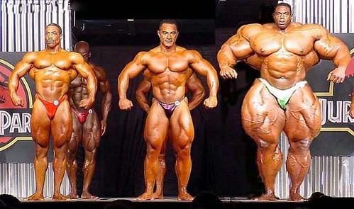
Body dysmorphic disorder – being excessively concerned and preoccupied with an imagined or minor defect in their appearance
http://farm1.static.flickr.com/144/367772579_ab90abdfd2.jpg

12.5 Homeostasis

Both muscular and skeletal systems work to maintain homeostasis.
Movement: Muscles move and are supported by skeleton
Protection: Both protect body parts
Bones are influenced by hormones to store and release calcium. Muscles need calcium for contractions.
Bones produce blood cells. Red blood cells carry oxygen needed to produce ATP by aerobic cellular respiration. White cells fight infection.
Muscles help regulate body temperature. Contracting muscles produce heat. That is why when you’re very cold, you shiver.


skyview.billings.k12.mt.us/.../shivering.gif

Exercise, Is the Concept Working?


http://supplementalscience.files.wordpress.com/2007/05/exercise.gif

I think we have intellectual awareness of the need to exercise. I’m certain we’ve all heard the call, seen the dismal statistics of obese-related health problems . . . and their cost. The estimated total cost of obesity in the United States in 2000 was about $117 billion, that was eight years ago.

I would say we all know we should be doing it. So why aren’t we? Here’s some sobering stats:

“Since the mid-seventies, the prevalence of overweight and obesity has increased sharply for both adults and children. Data from two NHANES surveys show that among adults aged 20–74 years the prevalence of obesity increased from 15.0% (in the 1976–1980 survey) to 32.9% (in the 2003–2004 survey). The two surveys also show increases in overweight among children and teens. For children aged 2–5 years, the prevalence of overweight increased from 5.0% to 13.9%; for those aged 6–11 years, prevalence increased from 6.5% to 18.8%; and for those aged 12–19 years, prevalence increased from 5.0% to 17.4%.”
“Despite the proven benefits of physical activity, more than 50% of U.S. adults do not get enough physical activity to provide health benefits; 24% are not active at all in their leisure time. Activity decreases with age, and sufficient activity is less common among women than men and among those with lower
incomes and less education. Insufficient physical activity is not
limited to adults. About two-thirds of young people in grades 9–12 are not engaged in recommended levels of physical activity. Daily participation in high school physical education classes dropped from 42% in 1991 to 33% in 2005.” http://www.cdc.gov/nccdphp/publications/aag/dnpa.htm


http://www.aafp.org/fpr/20001100/8_f1.jpg

Below is a map showing the growing rate of obesity in the USA over the last 15 years.

http://www.cdc.gov/nccdphp/publications/aag/dnpa.htm

I know the things in my world that make exercising difficult. It’s inconvenient, uncomfortable, sweaty, boring, and painful. Now, I realize we don’t have to lift weights to be healthy, and I don’t. There are far less demanding ways to lead an active life without becoming an athlete. The American Diabetes Association has several wonderful suggestions for gently incorporating movement into your day. Even just cleaning your house is a wonderful low-level activity. Getting up to change the channels and mowing the lawn are also suggested. Here’s a good one, playing with your kids.
This brings me to the “easy way”. We are creatures of comfort and it is hard to motivate ourselves to work harder than we have to. Most of our economy is based on convenience items: remotes for our TV’s, automobiles for everyone of age, elevators, even men in golf carts to drive you to the big building so you don’t have to walk too far. I’ve seen easy chairs with cup holders and meal trays built right in. Even our food is easier to prepare. Scissors are now a staple in my kitchen so I can cut those bags of frozen dinners open – seemed homemade since I dirtied a pan.
There are so many benefits to exercising regularly:
Physical activity improves quality of life.
Physical activity extends longevity, protects against the development of
CHD, stroke, hypertension, obesity, non-insulin-dependent diabetes
mellitus, osteoporosis, colon cancer, and depression.
Physical activity helps maintain full functioning and independence among
the elderly.
Relationships between physical activity and improved health include:
A reduced oxygen demand at any given level of physical activity.
A reduced tendency for blood to form clots where arteries have narrowed.
An increased elasticity in the arteries.
Changes in the brain and brain chemistry that may improve mood and
cognitive functioning.
More than 60% of US adults do not engage in the recommended amount
of activity.
Approximately 25% of US adults are not active at all.
A 1993 study concerning causes of death in the US: #1 Tobacco, #2 Lack of
exercise/poor diet, #3 Alcohol.
Chronic disease costs the US $655 Billion in healthcare costs.
In this year, 1.5 million people will have a heart attack in the US and
500,000 will die. http://www2.gsu.edu/~wwwfit/physicalactivity.html

Gamers now have video games that involve whole body movement, Wii is one. It’s quite popular at the nursing home I work at. With that game, people are moving as though they are playing a sport while holding a little box. This appears as a sport on the TV set and their movements are mimicked by the cartoon character that is playing them. Well, this is a start.
How do we get motivated? Peer pressure might help. Through government programs, involved citizens, and media coverage, many people might think again about getting off their you-know-whats. Scotland has many successful programs for activities. The constant for all of the programs is hiring enthusiastic, dedicated individuals to run them. Enthusiasm is contagious. In smaller communities, scheduled walking outings are offered a few times a week and locals need only to find the one that fits their schedule.

Encouragement and availability are key factors to participation. Of course, our food chain needs re-vamping as well, but that was our essay in the last unit.

Unit III Lab Project: Build a Movable Limb

Build a Limb Project

For this project, I will demonstrate how muscles and bones work together to produce movement. For this demo, I will model the arm.

Some raw materials:


The backscratch gave his life . . .


The operation: in progress.


After careful planning and assembling, the patient experiences - movement!

An explanation:

The motor neuron (white balloon in the hand of the operator/surgeon) signals the bicep. You can see the muscle responding to the stimuli by contracting. As it contracts, the diameter of the muscle increases, as mass remains the same (is it becomes shorter, it must get larger somewhere else). If this were more anatomically correct, the muscle would also be harder during contraction. The shortening of the muscle and pulling draws the forearm upwards, pivoting on the elbow joint.
Some stills . . .


Close up of motor unit


It's hard to see the action potential coarsing along the axon, there is a slight purple color to indicate that between the schwann cells. This potential will result in a release of ACh from the axon terminals. The neurotransmitter will then bind to the receptors in the sarcolemma. Then calcium will be released from the sarcoplasmic reticulum and will bind to troponin on the actin filament.


This deposition of calcium causes the troponin to move and expose the binding sites for the myosin.

Relaxed state of sarcomere


The myosin has heads that will extend to the binding sites on the actin forming a cross-bridge and will pull the actin closer together. ADP is utilized for this action and ATP is needed for the heads to release from the binding sites.
Contracted state of sarcomere

All of these actions happen at an amazing rate. Just think about how often you move without even thinking about it. Knowing the mechanics and chemical reactions needed to move one fibril makes my head spin. Even as I'm typing this report, I am amazed (can't think of a better word) by the effortless motions of my fingers in leiu of the magnitude of activities that are needed to accomplish it.

Movement Lab

Movement Lab

This is an attempt to explain how temperature and fatigue affects the function of muscle. This is important so we know what is needed to optimize our performance.


1. What are the three changes you observed in a muscle while it is working (contracted)?


The muscle shortened.

The muscle diameter enlarged.

The muscle became hard.


2. What effect did the cold temperature have on the action of your hand muscles? Explain.


I imagine that most people had a greater difference. My hands are always cold (Raynauld's Phenomenon). The cold water caused the muscles to ache and respond slowly.


3. In Figure 3, make a line graph of your results of the fatigue experiment. Be sure to fill in the values on the vertical axis.


Number of trials indicated above

Maybe the reps leveled off because my hand was warming up from exercising.


4. What effect did fatigue have on the action of your hand muscles? Explain.


I quickly felt the aching and slowing of response to continous squeezing. Initially the Creatine Phosphate pathway was utilized by the muscles, but this is good for only 5 seconds. Next, the muscles obtain ATP by fermentation, which produces a build-up of lactate. This is felt as aching in your muscles and is an anaerobic metabolism. I believe that the process was not able to keep up with the demand and that is why I could not maintain faster repetitions.


Below is an interesting chart showing how our muscles use the energy stores and methods.


Wednesday, April 2, 2008

Compendium: Chapters 13-14

Chapter 13: Nervous System

13.1 Overview of the Nervous System
Consists of:
Central Nervous System: brain and spinal cord
Peripheral Nervous System: nerves
Three functions:
Receives sensory input: internal and external
Integration: all inputs reviewed simultaneously
Generates motor output: stimulates muscles and glands
Nervous Tissue is made up of 2 types of cells:
Neurons: transmit signals within the nervous system
Three types:
Sensory Neuron: transfers messages from receptors to the Central Nervous System
Sensory Receptors: detects changes in the environment
Interneuron: located in the CNS and receives messages from the sensory neurons.
Motor Neuron: transmits from the CNS and generates motor and gland responses
Neuroglia: support and nourish neurons
Neurons have three parts:
Cell body: contains the nucleus and organelles
Dendrites: filigree-like extensions that receive signals from receptors or other neurons
Axon: conducts impulses
The Myelin Sheath is a protective covering along long axons of nerve cells. These are impartial coverings with gaps called the nodes of Ranvier.
Anatomy of a nerve
Interestingly, gray matter in the CNS is gray because the axons are not myelinated and white matter is. The myelin sheath protects and provides a passageway for regeneration if a nerve is severed. Multiple sclerosis and leukodystophries are disorders affecting the myelin sheath.
The nerve impulses are an exchange of ionic concentrations across the axon’s membrane. Sodium-potassium pumps maintain a potassium concentration greater than sodium within the cell giving it polarization and it is at its resting potential. When a stimulis is generated, the polarity changes along the course of the axon from node to node and the message is relayed along the PNS to the CNS and back to the muscles or glands. This change in polarity is called action potential. Damaged or unmyelinated axons move the impulse much slower. The intensity of pain or feeling depends on how often the nerve impulses are given in a short span of time. As soon as the impulse has passed, the ionic displacement returns to the resting potential because of repolarization.
The synapses are gaps that separate the sending from receiving neurons. Neurotransmitters are molecules stored in synaptic vessels that help transmit signals across these gaps. There are over 100 substances that have that capability. Here’s a sampling of just a few and what they effect:
Acetylcholine - voluntary movement of the muscles
Norepinephrine - wakefulness or arousal
Dopamine - voluntary movement and motivation, "wanting", pleasure, associated with addiction and love
Serotonin - memory, emotions, wakefulness, sleep and temperature regulation
GABA - inhibition of motor neurons
Glycine - spinal reflexes and motor behaviour
Neuromodulators - sensory transmission - especially pain
http://en.wikipedia.org/wiki/Neurotransmitter
A neuron has many different synapses with other neurons. Some are excitatory, other inhibitory. The response will depend on which signals the most.

13.2 The Central Nervous System

The spinal cord and the brain comprise the CNS. Meninges are membranes that protect the brain and spinal cord. Cerebrospinal fluid protects the brain and spinal cord as it fills the spaces in between the meninges and fills the ventricles of the brain. Inflammation of the meninges is meningitis.
There are four ventricles which are reservoirs for Cerebrospinal fluid. When these ventricles are blocked from draining, increased pressure can cause brain damage.

http://www.medicalook.com/systems_images/Ventricles_of_the_Brain.gif
White matter and gray matter make up the CNS.
The spinal cord originates from the base of the brain and extends down the vertebral canal. The nerves from the spinal cord extend from the cord through the openings between the vertebrae. There are softer discs which separate the vertebrae, when they slip, compression of a nerve may result in pain. Gray matter is the very core of the spinal cord which is surrounded by white matter. Dorsal and ventral nerves branch off the spinal cord and will join to form the spinal nerve which is part of the PNS.
All communication between the brain and PNS must transverse via the spinal cord. It is theorized that “gates” in the spinal cord regulate the flow of pain messages to the brain.
Severance of the spinal cord will “disengage” the functions and sensations of the body whose signals travel through that level in the cord. For example, a thoracic injury could render someone paralyzed and if the severance was higher the arms may be involved as well.
Some signals “arc” in the spinal cord and these responses are immediate and involuntary. We call these reflexes.
Lobes of a cerebral hemisphere

http://www.pennhealth.com/health_info/body_guide/reftext/images/anatomy_brain.jpg
The brain is separated into three sections.
The Cerebrum is the largest section and is divided into two hemispheres. These are distinctly divided by a deep fissure running from front to back along the top of the head. The cerebrum is divided into lobes (see image for location). The cerebral cortex is a covers the cerebral hemispheres with gray matter and has certain functions according to region:
Frontal: It is concerned with emotions, reasoning, planning, movement, and parts of speech. It is also involved in purposeful acts such as creativity, judgment, problem solving, and planning. Movement of the hand takes up a large portion of this motor area.
Parietal: Processes nerve impulses related to the senses, such as touch, pain, taste, pressure, and temperature. They also have language functions.
Temporal: The temporal lobes are responsible for hearing, memory, meaning, and language. They also play a role in emotion and learning. The temporal lobes are concerned with interpreting and processing auditory stimuli.
Occipital: The occipital lobe is involved with the brain's ability to recognize objects. It is responsible for our vision. http://library.thinkquest.org/J002391/functions.html
The white matter is tissue through which messages are sent from different areas of gray matter.
The Diencephalon is located centrally in the brain around the third ventricle. There are three regions within:
The hypothalamus regulates hunger, sleep, thirst, body temperature and water balance.
The thalamus receives all sensory inputs except smell and sends it to corresponding areas of the cerebrum.
The pineal gland secretes melatonin which regulates sleep rhythms and may also regulate the onset of puberty.


Perhaps result of hyperactive pineal gland . . .


The cerebellum is comprised mostly of white matter surrounded by gray matter. It is separated from the brain stem by the fourth ventricle. It receives input regarding the position of body parts and from the cerebral cortex about where these parts should be. After integrating this information, it will send motor impulses so we maintain balance and posture.
The brain stem is composed of four things:
Mid brain: transfers information between the cerebrum and spinal cord and some reflexes.
Pons: means “bridge” and transfers information between cerebellum and the rest of the CNS. It also has some reflex responses and regulates the rate of breathing.
Medulla Oblongata: maintains several reflexes affecting coughing, vomiting, sneezing, hiccupping, swallowing, and heartbeat, breathing and vasoconstriction.
Reticular Formation: (lines the brain stem) keeps you awake and will react to startling sounds/events. Damage to this area will render one comatose.

13.3 The Limbic System and Higher Mental Functions

Limbic system structures are involved in many of our emotions and motivations, particularly those that are related to survival. Such emotions include fear, anger, and emotions related to sexual behavior. The limbic system is also involved in feelings of pleasure that are related to our survival, such as those experienced from eating and sex. Certain structures of the limbic system are involved in memory as well. http://biology.about.com/od/anatomy/a/aa042205a.htm

The amygdale lends emotional overtones to our experiences. The fight or fright response is initiated by the amydgala. This response can be overridden by the frontal cortex from which we have judgment. Old memories can elicit emotional responses because of the amydgala.
The hippocampus helps us learn and remember.
We have different types of memory:
Short-term: originates in the pre-frontal region.
Long-term: a mixture of facts (semantic) and people or events (episodic). These memories are stored in the areas of the brain where the events originated.
Skill memory: repetitive activities become memorized.

13.4 The Peripheral Nervous System

The peripheral nervous system is the nervous system outside of the CNS. We have 12 pairs of cranial nerves that originate from the brain and are primarily concerned with the neck, face and head. There is one nerve, the vagus nerve, which runs to nearly all the internal organs as well as the throat. The spinal nerves are paired from both sides of the spinal cord. They are mixed nerves containing both sensory and motor fibers. Their placement determines which region of the body they serve.
The somatic system includes all nerves controlling the muscular system and external sensory receptors. The reflex arc is the pathway from the sensory receptors to the spinal cord to the motor neuron to the effector which elicits an immediate response without thinking about it. Your brain also receives the signal only to perceive the pain or stimulus.
The autonomic system is composed of the sympathetic and parasympathetic divisions.
The sympathetic system is the pathway for fight or flight responses. It increases heart rate and dilates the bronchi and shuts down the digestive tract. We don’t need that to rob us of energy for the crisis we’re in.
The parasympathetic system promotes relaxation and digestion.
Sympathetic and parasympathetic systems

http://www.ptsdforum.org/images/autonomic-system.gif

13.5 Drug Abuse

Drugs used for altered perceptions and feelings affect either our limbic system or promote or decrease the action of some neurotransmitters. A stimulant will excite neurons and depressants will diminish the likelihood of excitation. We have a reward circuit in our brain that equates activities such as eating as being pleasurable. Substance abusers will take drugs to experience this feeling and even neglect legitimate physical requirements. Addictions can be psychological or physical.
Alcohol is the most common and most accepted drug. It crosses cell membranes and damages several tissues. Liver damage is incurred from chronically removing this toxin. Alcohol is a depressant and will affect coordination, lead to a feeling of relaxation, a lowering of inhibitions, slurred speech and possibly vomiting. Coma or death can occur when ETOH levels are too high. Chronic consumption causes brain damage, decreases brain size and increase the size of the ventricles.
Nicotine is a stimulant. It increases skeletal muscle activity, elevates heart rate and BP and peristalsis. Nicotine is a physical and psychological addiction. 80-90% of smokers will fail at quitting.
Cocaine is a stimulant that interferes with the re-uptake of dopamine at synapses. Users are treated to a rush of well-being lasting up to 30 minutes. Between binges, one may experience fatigue and irritability. Crack is a form of cocaine that is smoked. This brings a quicker, more intense high.
Methamphetamine is a synthetic, easily made drug. It is often made in home-based labs. This is also available in two forms – powder and crystal (smoked). It is a stimulant and will elevate the mood of the user. Some people will become violent if they become agitated. Chronic use may result in paranoia and hallucinations. Ecstasy is similar to Meth but will not cause hallucinations.
Heroin comes from poppies. It is a depressant and an opiate and has pain-killing effects also delivering a rush and euphoria. It can be snorted, smoked or injected and is the most abused opiate. Opiates cloud thinking, trigger the reward circuit, and can cause nausea and vomiting. Needle sharing is the primary source of hepatitis and HIV/AIDS. Over 300,000 people use heroin every year.
Marijuana comes from the cannabis sativa. It is common to consider the effects of marijuana as mild. Some euphoria may occur along with changes in judgment. Regular use can cause cravings. Heavy users may experience more severe reactions such as, hallucinations and paranoia. It is a psychoactive drug.
Loves his greens . . .
http://www.cosmosmagazine.com/

Chapter 14: Senses

14.1 Sensory Receptors and Sensations

These are dendrites that detect types of stimuli. Exteroceptors detect external stimuli and Interoceptors detect internal stimuli. There are different types of exteroceptors:
Chemoreceptors – taste and smell. Sensors in the carotids and aorta sense pH and adjustments are made if it is out of range.
Pain receptors – dendrites exposed to chemicals released by damaged cells.
Photoreceptor – react to light (eyes).
Mechanoreceptors – sense of touch. Pressure that is felt by gravity or sound waves (hair cells). Proprioceptors sense the position of our limbs.
Thermoreceptors – stimulated by changes in temperature.
These receptors will integrate all signals prior to sending. Sometimes adaptation will occur and you may notice a decrease sensation. This usually happens when the stimulis is chronic, such as an aroma which may be strong at first exposure, but will not be as noticeable after a while.

14.2 Proprioceptors and Cutaneous Receptors

Proprioceptors detect the position of our limbs. Cutaneous receptors are located in the epidermis and the dermis. These detect touch, pressure, pain and temperature. Internal organs have pain receptors (nociceptors) that are stimulated by chemicals released from damaged cells. Interestly, Aspirin inhibits the synthesis of one type of these chemicals.

14.3 Senses of Taste and Smell

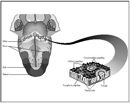
We have approximately 3,000 taste buds located mostly on our tongue. There are a few scattered about on our hard palate, pharynx, and epiglottis. Different areas on our tongue detect different types of tastes.
Areas of tastes and anatomy of the tongue's surface


What we actually taste is a result of the cortex combining all the flavors and smells to give the interpretation. 80-90% of what we taste is attributed to what we smell. Olfactory cells are located high in the nasal cavity. These are protein receptors and react to certain molecules. Our sense of smell is also connected to our limbic system and that is why certain smells trigger memories and emotional responses.
As we age, these cells decrease in number and sensitivity.

14.4 Sense of Vision
Here's lookin' at you, kid.

blogofunny.blogspot.com/2007/07/eyesight.html

The eyeball is a globe whose outer mantel is composed of 3 layers. The sclera (white of the eye), Choroid (vascular and absorbs light), and the retina (contains sensory receptors).
In the front of the eye, the sclera becomes the cornea or the window of the eye. The Choroid becomes the iris, the colored part of your eye which regulates how much light enters the eyeball. The choroid also is behind the iris and will change the shape of the lens for focusing. This lens separates the anterior and posterior compartments. The aqueous humor is a liquid which fills the chamber of the anterior eye. The pressure of this fluid is regulated via ducts and when these ducts are blocked, increased pressure can cause nerve damage. This is called glaucoma. The gelatinous fluid in the posterior chamber is called vitreous humor. The retina has rod (light sensitive) and cone cells (color sensitive). We have up to 150 million rod cells and 6 million cone cells. The stimuli from these cells pass to bipolar and ganglion cells and then along the optic nerve to the visual cortex where more integration occurs. We have a blind spot in each eye which occurs where the optic nerve exits the eye. These are not the same location for both eyes and therefore, it is not noticeable when both eyes are open.
Misshapened eyeballs alter vision. Elongated ones cause distance to become blurry and shortened ones cause near objects to be blurry. Some are uneven causing astigmatism.

14.5 Sense of Hearing


The pinna is the part of the ear we see. Inside are several structures that contribute to hearing and balance. Sweat glands in the upper wall of the canal produce wax which reduces the amount of substances that enter our ear. The ear drum (tympanic membrane) is the beginning of the middle ear and ends at the round window. The inner ear is fluid-filled. Sound waves enter the auditory canal and their slight vibration causes the tympanic membrane to vibrate. This movement is picked up the malleus, incus and stapes which will amplify the signal 20X. The stapes will strike the membrane over the oval window and this vibration will be passed to the fluid in the cochlea. Little hairs in the cochlea will bend and send signals along the cochlear nerve to the auditory cortex. Loud noises cause increased stimulation.

14.6 Sense of Equilibrium
Location of Cupula in Semicircular Canal


http://mems.eng.uci.edu/Personnel/jiayin/main.h3.jpg

Mechanoreceptors in the semicircular canals detect motion. The cupula will bend as fluid is displaced by movement and receptors signal the change of position. The utricle and saccule contain little hair cells which will bend with head movements as well. If we suddenly stop a dizzying ride, the cupula move slowly and our brain still thinks we’re spinning when our eyes clearly show we are not, causing dizziness.
When do we get too old for swings?

http://www.balanceandmobility.com/patient_info/images/8.jpg