Chapter 13: Nervous System
13.1 Overview of the Nervous System
Consists of:
Central Nervous System: brain and spinal cord
Peripheral Nervous System: nerves
Three functions:
Receives sensory input: internal and external
Integration: all inputs reviewed simultaneously
Generates motor output: stimulates muscles and glands
Nervous Tissue is made up of 2 types of cells:
Neurons: transmit signals within the nervous system
Three types:
Sensory Neuron: transfers messages from receptors to the Central Nervous System
Sensory Receptors: detects changes in the environment
Interneuron: located in the CNS and receives messages from the sensory neurons.
Motor Neuron: transmits from the CNS and generates motor and gland responses
Neuroglia: support and nourish neurons
Neurons have three parts:
Cell body: contains the nucleus and organelles
Dendrites: filigree-like extensions that receive signals from receptors or other neurons
Axon: conducts impulses
The Myelin Sheath is a protective covering along long axons of nerve cells. These are impartial coverings with gaps called the nodes of Ranvier.
Anatomy of a nerve

Interestingly, gray matter in the CNS is gray because the axons are not myelinated and white matter is. The myelin sheath protects and provides a passageway for regeneration if a nerve is severed. Multiple sclerosis and leukodystophries are disorders affecting the myelin sheath.
The nerve impulses are an exchange of ionic concentrations across the axon’s membrane. Sodium-potassium pumps maintain a potassium concentration greater than sodium within the cell giving it polarization and it is at its resting potential. When a stimulis is generated, the polarity changes along the course of the axon from node to node and the message is relayed along the PNS to the CNS and back to the muscles or glands. This change in polarity is called action potential. Damaged or unmyelinated axons move the impulse much slower. The intensity of pain or feeling depends on how often the nerve impulses are given in a short span of time. As soon as the impulse has passed, the ionic displacement returns to the resting potential because of repolarization.
The synapses are gaps that separate the sending from receiving neurons. Neurotransmitters are molecules stored in synaptic vessels that help transmit signals across these gaps. There are over 100 substances that have that capability. Here’s a sampling of just a few and what they effect:
Acetylcholine - voluntary movement of the muscles
Norepinephrine - wakefulness or arousal
Dopamine - voluntary movement and motivation, "wanting", pleasure, associated with addiction and love
Serotonin - memory, emotions, wakefulness, sleep and temperature regulation
GABA - inhibition of motor neurons
Glycine - spinal reflexes and motor behaviour
Neuromodulators - sensory transmission - especially pain
http://en.wikipedia.org/wiki/Neurotransmitter
A neuron has many different synapses with other neurons. Some are excitatory, other inhibitory. The response will depend on which signals the most.
13.2 The Central Nervous System
The spinal cord and the brain comprise the CNS. Meninges are membranes that protect the brain and spinal cord. Cerebrospinal fluid protects the brain and spinal cord as it fills the spaces in between the meninges and fills the ventricles of the brain. Inflammation of the meninges is meningitis.
There are four ventricles which are reservoirs for Cerebrospinal fluid. When these ventricles are blocked from draining, increased pressure can cause brain damage.

http://www.medicalook.com/systems_images/Ventricles_of_the_Brain.gif
White matter and gray matter make up the CNS.
The spinal cord originates from the base of the brain and extends down the vertebral canal. The nerves from the spinal cord extend from the cord through the openings between the vertebrae. There are softer discs which separate the vertebrae, when they slip, compression of a nerve may result in pain. Gray matter is the very core of the spinal cord which is surrounded by white matter. Dorsal and ventral nerves branch off the spinal cord and will join to form the spinal nerve which is part of the PNS.
All communication between the brain and PNS must transverse via the spinal cord. It is theorized that “gates” in the spinal cord regulate the flow of pain messages to the brain.
Severance of the spinal cord will “disengage” the functions and sensations of the body whose signals travel through that level in the cord. For example, a thoracic injury could render someone paralyzed and if the severance was higher the arms may be involved as well.
Some signals “arc” in the spinal cord and these responses are immediate and involuntary. We call these reflexes.
Lobes of a cerebral hemisphere

http://www.pennhealth.com/health_info/body_guide/reftext/images/anatomy_brain.jpg
The brain is separated into three sections.
The Cerebrum is the largest section and is divided into two hemispheres. These are distinctly divided by a deep fissure running from front to back along the top of the head. The cerebrum is divided into lobes (see image for location). The cerebral cortex is a covers the cerebral hemispheres with gray matter and has certain functions according to region:
Frontal: It is concerned with emotions, reasoning, planning, movement, and parts of speech. It is also involved in purposeful acts such as creativity, judgment, problem solving, and planning. Movement of the hand takes up a large portion of this motor area.
Parietal: Processes nerve impulses related to the senses, such as touch, pain, taste, pressure, and temperature. They also have language functions.
Temporal: The temporal lobes are responsible for hearing, memory, meaning, and language. They also play a role in emotion and learning. The temporal lobes are concerned with interpreting and processing auditory stimuli.
Occipital: The occipital lobe is involved with the brain's ability to recognize objects. It is responsible for our vision. http://library.thinkquest.org/J002391/functions.html
The white matter is tissue through which messages are sent from different areas of gray matter.
The Diencephalon is located centrally in the brain around the third ventricle. There are three regions within:
The hypothalamus regulates hunger, sleep, thirst, body temperature and water balance.
The thalamus receives all sensory inputs except smell and sends it to corresponding areas of the cerebrum.
The pineal gland secretes melatonin which regulates sleep rhythms and may also regulate the onset of puberty.
Perhaps result of hyperactive pineal gland . . .

The cerebellum is comprised mostly of white matter surrounded by gray matter. It is separated from the brain stem by the fourth ventricle. It receives input regarding the position of body parts and from the cerebral cortex about where these parts should be. After integrating this information, it will send motor impulses so we maintain balance and posture.
The brain stem is composed of four things:
Mid brain: transfers information between the cerebrum and spinal cord and some reflexes.
Pons: means “bridge” and transfers information between cerebellum and the rest of the CNS. It also has some reflex responses and regulates the rate of breathing.
Medulla Oblongata: maintains several reflexes affecting coughing, vomiting, sneezing, hiccupping, swallowing, and heartbeat, breathing and vasoconstriction.
Reticular Formation: (lines the brain stem) keeps you awake and will react to startling sounds/events. Damage to this area will render one comatose.
13.3 The Limbic System and Higher Mental Functions
Limbic system structures are involved in many of our emotions and motivations, particularly those that are related to survival. Such emotions include fear, anger, and emotions related to sexual behavior. The limbic system is also involved in feelings of pleasure that are related to our survival, such as those experienced from eating and sex. Certain structures of the limbic system are involved in memory as well. http://biology.about.com/od/anatomy/a/aa042205a.htm
The amygdale lends emotional overtones to our experiences. The fight or fright response is initiated by the amydgala. This response can be overridden by the frontal cortex from which we have judgment. Old memories can elicit emotional responses because of the amydgala.
The hippocampus helps us learn and remember.
We have different types of memory:
Short-term: originates in the pre-frontal region.
Long-term: a mixture of facts (semantic) and people or events (episodic). These memories are stored in the areas of the brain where the events originated.
Skill memory: repetitive activities become memorized.
13.4 The Peripheral Nervous System
The peripheral nervous system is the nervous system outside of the CNS. We have 12 pairs of cranial nerves that originate from the brain and are primarily concerned with the neck, face and head. There is one nerve, the vagus nerve, which runs to nearly all the internal organs as well as the throat. The spinal nerves are paired from both sides of the spinal cord. They are mixed nerves containing both sensory and motor fibers. Their placement determines which region of the body they serve.
The somatic system includes all nerves controlling the muscular system and external sensory receptors. The reflex arc is the pathway from the sensory receptors to the spinal cord to the motor neuron to the effector which elicits an immediate response without thinking about it. Your brain also receives the signal only to perceive the pain or stimulus.
The autonomic system is composed of the sympathetic and parasympathetic divisions.
The sympathetic system is the pathway for fight or flight responses. It increases heart rate and dilates the bronchi and shuts down the digestive tract. We don’t need that to rob us of energy for the crisis we’re in.
The parasympathetic system promotes relaxation and digestion.
Sympathetic and parasympathetic systems

http://www.ptsdforum.org/images/autonomic-system.gif
13.5 Drug Abuse
Drugs used for altered perceptions and feelings affect either our limbic system or promote or decrease the action of some neurotransmitters. A stimulant will excite neurons and depressants will diminish the likelihood of excitation. We have a reward circuit in our brain that equates activities such as eating as being pleasurable. Substance abusers will take drugs to experience this feeling and even neglect legitimate physical requirements. Addictions can be psychological or physical.
Alcohol is the most common and most accepted drug. It crosses cell membranes and damages several tissues. Liver damage is incurred from chronically removing this toxin. Alcohol is a depressant and will affect coordination, lead to a feeling of relaxation, a lowering of inhibitions, slurred speech and possibly vomiting. Coma or death can occur when ETOH levels are too high. Chronic consumption causes brain damage, decreases brain size and increase the size of the ventricles.
Nicotine is a stimulant. It increases skeletal muscle activity, elevates heart rate and BP and peristalsis. Nicotine is a physical and psychological addiction. 80-90% of smokers will fail at quitting.
Cocaine is a stimulant that interferes with the re-uptake of dopamine at synapses. Users are treated to a rush of well-being lasting up to 30 minutes. Between binges, one may experience fatigue and irritability. Crack is a form of cocaine that is smoked. This brings a quicker, more intense high.
Methamphetamine is a synthetic, easily made drug. It is often made in home-based labs. This is also available in two forms – powder and crystal (smoked). It is a stimulant and will elevate the mood of the user. Some people will become violent if they become agitated. Chronic use may result in paranoia and hallucinations. Ecstasy is similar to Meth but will not cause hallucinations.
Heroin comes from poppies. It is a depressant and an opiate and has pain-killing effects also delivering a rush and euphoria. It can be snorted, smoked or injected and is the most abused opiate. Opiates cloud thinking, trigger the reward circuit, and can cause nausea and vomiting. Needle sharing is the primary source of hepatitis and HIV/AIDS. Over 300,000 people use heroin every year.
Marijuana comes from the cannabis sativa. It is common to consider the effects of marijuana as mild. Some euphoria may occur along with changes in judgment. Regular use can cause cravings. Heavy users may experience more severe reactions such as, hallucinations and paranoia. It is a psychoactive drug.
Loves his greens . . .

http://www.cosmosmagazine.com/
Chapter 14: Senses
14.1 Sensory Receptors and Sensations
These are dendrites that detect types of stimuli. Exteroceptors detect external stimuli and Interoceptors detect internal stimuli. There are different types of exteroceptors:
Chemoreceptors – taste and smell. Sensors in the carotids and aorta sense pH and adjustments are made if it is out of range.
Pain receptors – dendrites exposed to chemicals released by damaged cells.
Photoreceptor – react to light (eyes).
Mechanoreceptors – sense of touch. Pressure that is felt by gravity or sound waves (hair cells). Proprioceptors sense the position of our limbs.
Thermoreceptors – stimulated by changes in temperature.
These receptors will integrate all signals prior to sending. Sometimes adaptation will occur and you may notice a decrease sensation. This usually happens when the stimulis is chronic, such as an aroma which may be strong at first exposure, but will not be as noticeable after a while.
14.2 Proprioceptors and Cutaneous Receptors
Proprioceptors detect the position of our limbs. Cutaneous receptors are located in the epidermis and the dermis. These detect touch, pressure, pain and temperature. Internal organs have pain receptors (nociceptors) that are stimulated by chemicals released from damaged cells. Interestly, Aspirin inhibits the synthesis of one type of these chemicals.
14.3 Senses of Taste and Smell
We have approximately 3,000 taste buds located mostly on our tongue. There are a few scattered about on our hard palate, pharynx, and epiglottis. Different areas on our tongue detect different types of tastes.
Chapter 14: Senses
14.1 Sensory Receptors and Sensations
These are dendrites that detect types of stimuli. Exteroceptors detect external stimuli and Interoceptors detect internal stimuli. There are different types of exteroceptors:
Chemoreceptors – taste and smell. Sensors in the carotids and aorta sense pH and adjustments are made if it is out of range.
Pain receptors – dendrites exposed to chemicals released by damaged cells.
Photoreceptor – react to light (eyes).
Mechanoreceptors – sense of touch. Pressure that is felt by gravity or sound waves (hair cells). Proprioceptors sense the position of our limbs.
Thermoreceptors – stimulated by changes in temperature.
These receptors will integrate all signals prior to sending. Sometimes adaptation will occur and you may notice a decrease sensation. This usually happens when the stimulis is chronic, such as an aroma which may be strong at first exposure, but will not be as noticeable after a while.
14.2 Proprioceptors and Cutaneous Receptors
Proprioceptors detect the position of our limbs. Cutaneous receptors are located in the epidermis and the dermis. These detect touch, pressure, pain and temperature. Internal organs have pain receptors (nociceptors) that are stimulated by chemicals released from damaged cells. Interestly, Aspirin inhibits the synthesis of one type of these chemicals.
14.3 Senses of Taste and Smell
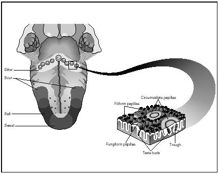
We have approximately 3,000 taste buds located mostly on our tongue. There are a few scattered about on our hard palate, pharynx, and epiglottis. Different areas on our tongue detect different types of tastes.
Areas of tastes and anatomy of the tongue's surface

What we actually taste is a result of the cortex combining all the flavors and smells to give the interpretation. 80-90% of what we taste is attributed to what we smell. Olfactory cells are located high in the nasal cavity. These are protein receptors and react to certain molecules. Our sense of smell is also connected to our limbic system and that is why certain smells trigger memories and emotional responses.
As we age, these cells decrease in number and sensitivity.
14.4 Sense of Vision
Here's lookin' at you, kid.

blogofunny.blogspot.com/2007/07/eyesight.html
The eyeball is a globe whose outer mantel is composed of 3 layers. The sclera (white of the eye), Choroid (vascular and absorbs light), and the retina (contains sensory receptors).
In the front of the eye, the sclera becomes the cornea or the window of the eye. The Choroid becomes the iris, the colored part of your eye which regulates how much light enters the eyeball. The choroid also is behind the iris and will change the shape of the lens for focusing. This lens separates the anterior and posterior compartments. The aqueous humor is a liquid which fills the chamber of the anterior eye. The pressure of this fluid is regulated via ducts and when these ducts are blocked, increased pressure can cause nerve damage. This is called glaucoma. The gelatinous fluid in the posterior chamber is called vitreous humor. The retina has rod (light sensitive) and cone cells (color sensitive). We have up to 150 million rod cells and 6 million cone cells. The stimuli from these cells pass to bipolar and ganglion cells and then along the optic nerve to the visual cortex where more integration occurs. We have a blind spot in each eye which occurs where the optic nerve exits the eye. These are not the same location for both eyes and therefore, it is not noticeable when both eyes are open.
Misshapened eyeballs alter vision. Elongated ones cause distance to become blurry and shortened ones cause near objects to be blurry. Some are uneven causing astigmatism.
14.5 Sense of Hearing

The pinna is the part of the ear we see. Inside are several structures that contribute to hearing and balance. Sweat glands in the upper wall of the canal produce wax which reduces the amount of substances that enter our ear. The ear drum (tympanic membrane) is the beginning of the middle ear and ends at the round window. The inner ear is fluid-filled. Sound waves enter the auditory canal and their slight vibration causes the tympanic membrane to vibrate. This movement is picked up the malleus, incus and stapes which will amplify the signal 20X. The stapes will strike the membrane over the oval window and this vibration will be passed to the fluid in the cochlea. Little hairs in the cochlea will bend and send signals along the cochlear nerve to the auditory cortex. Loud noises cause increased stimulation.
14.6 Sense of Equilibrium
Location of Cupula in Semicircular Canal

http://mems.eng.uci.edu/Personnel/jiayin/main.h3.jpg
Mechanoreceptors in the semicircular canals detect motion. The cupula will bend as fluid is displaced by movement and receptors signal the change of position. The utricle and saccule contain little hair cells which will bend with head movements as well. If we suddenly stop a dizzying ride, the cupula move slowly and our brain still thinks we’re spinning when our eyes clearly show we are not, causing dizziness.
When do we get too old for swings?

http://www.balanceandmobility.com/patient_info/images/8.jpg

http://www.balanceandmobility.com/patient_info/images/8.jpg

2 comments:
it was nice and i have learned a lot just maintain it..i love it very much...
it was nice and i love it so much......
Post a Comment